Publications
Group highlights
For a full list of publications and patents see below or go to Google Scholar.

We explore the potential for dual-energy CT imaging with joint megavoltage and kilovoltage x-ray spectra to alleviate metal artifacts and improve soft-tissue contrast.
G Jadick, G Schlafly, P La Rivière
Journal of Medical Imaging (2024)
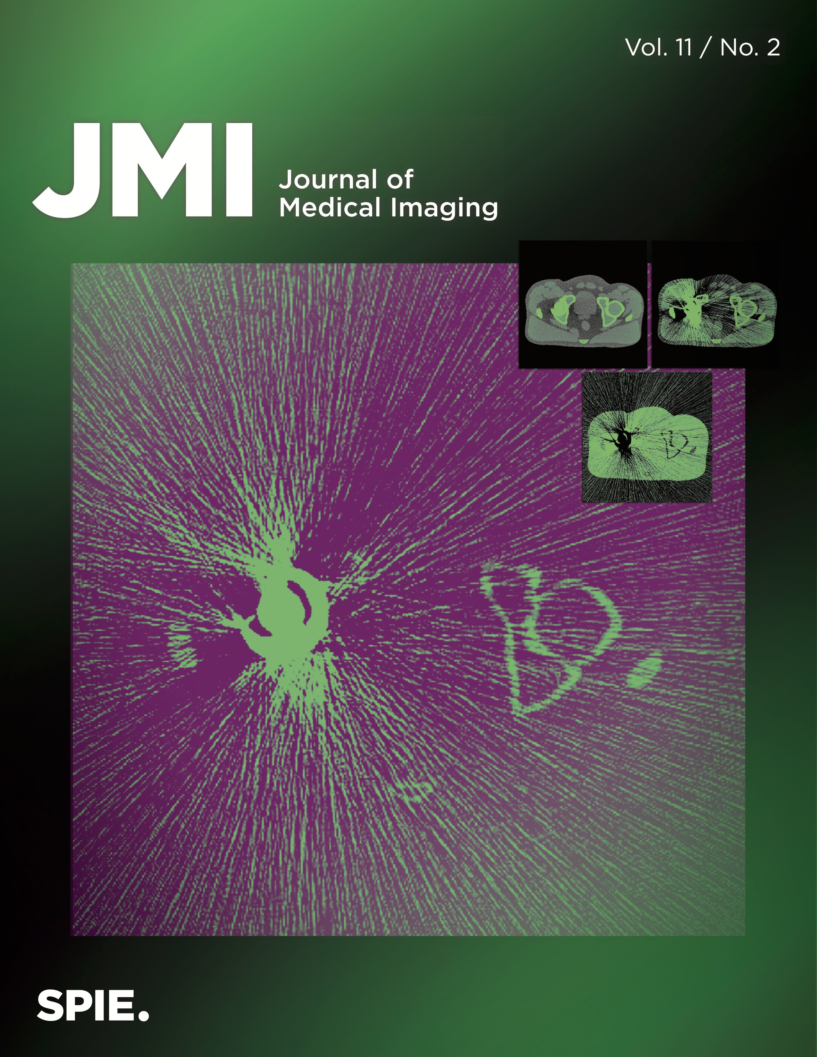
Our publication was featured on the cover of JMI, displaying severe metal artifacts in a simulated CT image due to a steel hip implant.

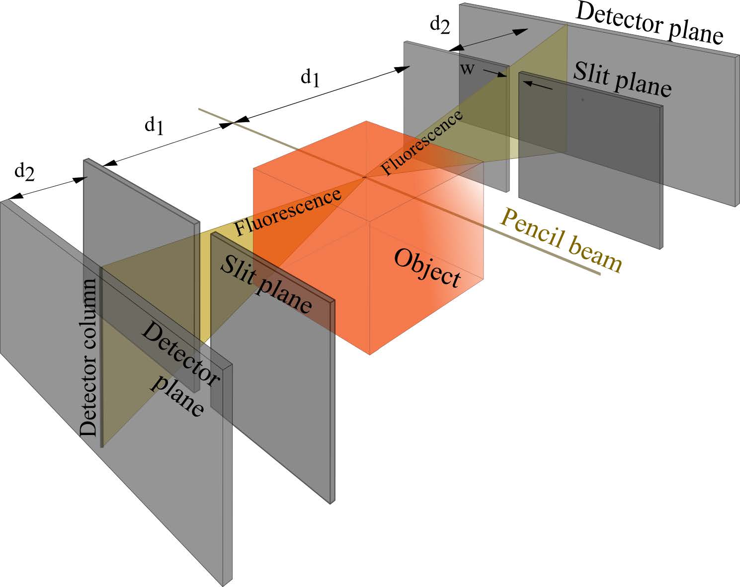
We investigate the joint estimation of metal and attenuation maps of a pencil-beam XFET system. Using simulated data, re demonstrate an accurate joint reconstruction.
H DeBrosse, T Chandler, LJ Meng, P La Rivière
IEEE trans. radiat. plasma med. sci. (2022)
X-ray fluorescence emission tomography (XFET) is an emerging imaging modality that images the spatial distribution of metal without requiring biochemical modification or radioactivity.

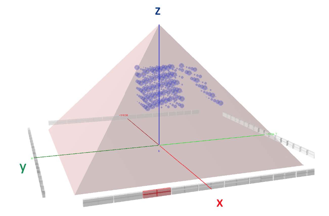
We plan to field a telescope system that has upwards of 100 times the sensitivity of the equipment that has recently been used at the Great Pyramid, will image muons from nearly all angles and will, for the first time, produce a true tomographic image of such a large structure.
A Bross, EC Dukes, R Ehrlich, E Fernandez, S Dukes, M Gobashy, I Jamieson, P La Rivière, M Liu, G Marouard, N Moeller, A Pla-Dalmau, P Rubinov, O Shohoud, Phil Vargas, T Welch
Patents
D Modgil, P La Rivière, Y Liu, Z Yu
Method and apparatus for spectral computed tomography (CT) with multi-material decomposition into three or more material components
US11238585B2 (2022)
H Shroff, Y Wu, X Han, P La Rivière
Systems and methods for producing isotropic in-plane super-resolution images from line-scanning confocal microscopy
WO2022150506A1 (2022)
H Frisch, E Angelico, P La Rivière, B Adams, E Spieglan, J Shida, A Elagin, K Domurat-Sousa
Positron emission tomography systems based on ionization-activated organic fluor molecules, planar pixelated photodetectors, or both
WO2022093732A1 (2022)
See more patents
List of publications since 2022
Last updated: July 2024
Vascular Plugs Improve Pulmonary Arteriovenous Malformation Occlusion over Coil Embolization Alone: A Proof-of-Concept Study using Dual Energy Computed Tomography
Q Yu, S Zangan, P La Rivière, L Landeras, B Funaki
Journal of Vascular and Interventional Radiology (2024)
A High-Sensitivity Benchtop X-Ray Fluorescence Emission Tomography (XFET) System With a Full-Ring of X-Ray Imaging-Spectrometers and a Compound-Eye Collimation Aperture
S Mandot, EM Zannoni, LiLng Cai, X Nie, P La Rivière, MD Wilson, LJ Meng
IEEE Transactions on Medical Imaging 43, 5 (2024)
Sinogram domain angular upsampling of sparse-view micro-CT with dense residual hierarchical transformer and attention-weighted loss
AS Adishesha, DJ Vanselow, P La Rivière, K Cheng and SX Huang.
Comput. Methods Prog. Biomed 242 C (2024)
Three-dimensional structured illumination microscopy with enhanced axial resolution
X Li, Y Wu, Y Su, I Rey-Suarez, C Matthaeus, TB Updegrove, Z Wei, L Zhang, H Sasaki, Y Li, M Guo, JP Giannini, HD Vishwasrao, J Chen, SJ Lee, L Shao, H Liu, KS Ramamurthi, JW Taraska, A Upadhyaya, P La Rivière, H Shroff
Nature Biotechnology 41, 9 (2023)
X-ray Fluorescence Emission Tomography of Implantable Scintillating Microparticles for X-Ray induced Optogenetic Application
S Mandot, EM Zannoni, P La Rivière, LJ Meng
Journal of Nuclear Medicine 64 (2023)
Phase-diversity-based wavefront sensing for fluorescence microscopy
C Johnson, M Guo, MC Schneider, Y Su, S Khuon, N Reiser, Y Wu, P La Rivière, H Shroff
Optica 11, 6 (2024)
Dual-energy computed tomography imaging with megavoltage and kilovoltage X-ray spectra
G Jadick, G Schlafly, P La Rivière
Journal of Medical Imaging (2024)
Effect of Detector Placement on Joint Estimation in X-ray Fluorescence Emission Tomography
H DeBrosse, LJ Meng, P La Rivière
IEEE trans. radiat. plasma med. sci. (2023)
Joint Estimation of Metal Density and Attenuation Maps with Pencil Beam XFET
H DeBrosse, T Chandler, LJ Meng, P La Rivière
IEEE trans. radiat. plasma med. sci. (2022)
Stopping Power Simulation for Use in Muon Tomography
I Jamieson, A Bross, EC Dukes, R Ehrlich, E Fernandez, S Dukes, M Gobashy, P La Rivière, G Marouard, N Moeller, A Pla-Dalmau, P Rubinov, O Shohoud, P Vargas, T Welch
JAIS 281 (2022)
Computerized Texture Analysis of Optical Coherence Tomography Angiography of Choriocapillaris in Normal Eyes of Young and Healthy Subjects
A Movahedan, P Vargas, J Moir, G Kaufmann, L Chun, C Smith, N Massamba, P La Rivière, D Skondra
Cells 11, 12 (2022)
A wide-field micro-computed tomography detector: micron resolution at half-centimetre scale
Maksim A Yakovlev, Daniel J Vanselow, Mee Siing Ngu, Carolyn R Zaino, Spencer R Katz, Yifu Ding, Dula Parkinson, Steve Yuxin Wang, Khai Chung Ang, Patrick La Rivière, Keith C Cheng
J Synchrotron Rad (2022)
Tomographic Muon Imaging of the Great Pyramid of Giza
A Bross, EC Dukes, R Ehrlich, E Fernandez, S Dukes, M Gobashy, I Jamieson, P La Rivière, M Liu, G Marouard, N Moeller, A Pla-Dalmau, P Rubinov, O Shohoud, Phil Vargas, T Welch
arXiv:2202.08184 (2022)
Synergistic checkpoint-blockade and radiotherapy–radiodynamic therapy via an immunomodulatory nanoscale metal-organic framework
K Ni, Z Xu, A Culbert, T Luo, N Guo, K Yang, E Pearson, B Preusser, T Wu, PLa Rivière, R Weichselbaum, M Spiotto, W Lin
Nat Biomed Eng 6, 2 (2022)
Uncovering the Effects of Symbiosis and Temperature on Coral Calcification
Zoe Dellaert, Phillip A Vargas, Patrick J La Rivière, Loretta M Roberson
The Biological Bulletin 242, 1 (2022)
Three-dimensional structured illumination microscopy with enhanced axial resolution
X Li, Y Wu, Y Su, I Suarez, C Matthaeus, T Updegrove, Z Wei, L Zhang, H Sasaki, Y Li, M Guo, J Giannini, H Vishwasrao, J Chen, SJ Lee, L Shao, H Liu, K Ramamurthi, J Taraska, A Upadhyaya, P La Rivière, H Shroff
biorxiv:10.1101/2022.07.20.500834 (2022)
Model-free analysis in the spectral domain of postmortem mouse brain EPSI reveals inconsistencies with model-based analyses of the free induction decay
Scott Trinkle, Gregg Wildenberg, Narayanan Kasthuri, Patrick La Rivière, Sean Foxley
biorxiv:10.1101/2022.02.24.481824 (2022)